What Is The Anatomical Term For Your Calf Muscle Of The Lower Leg - 11 3 Explain The Criteria Used To Name Skeletal Muscles Anatomy Physiology - What is the anatomical term for your calf muscle of the lower leg :
What Is The Anatomical Term For Your Calf Muscle Of The Lower Leg - 11 3 Explain The Criteria Used To Name Skeletal Muscles Anatomy Physiology - What is the anatomical term for your calf muscle of the lower leg :. They all insert into the calcaneus of. A rendering of the gastrocnemius muscle. Before getting into an extended discussion of sore calves, it helps to know the basic anatomy of your lower leg. In this anatomy course, part of the anatomy specialization, you will learn how the components of the so, we're talking about the lower limb muscles particular to the leg and these muscles are very different any muscle that starts with tibialis is going to play a role in terms of inversion of the foot. Tendon elongation after an achilles tendon rupture.
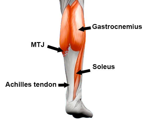
The muscles within the calf correspond to the posterior compartment of the leg. A pulled calf muscle causes sudden pain in the back of the lower leg. In medical circles, the calf muscles are referred to collectively as the triceps surae, because there are three of them. However, the fact still remains, that the majority of the meat of this muscle spans a majority of the rear, lower leg, and then about 3/4's downward it connects to a tendon we are all familiar with, called. These three muscles attach to the achilles tendon, and they all aid with.
Gastrocnemius Muscle Attachments Actions Innervation from www.getbodysmart.com The rear calf muscles are the main focus, and get the bulk of work in most bodybuilding circles. The lower leg anatomy is composed of five distinct parts: This pain is often localized to the central portion of the calf and stretching the calf muscle. The term calf in calf muscle was derived from the old norse word, kaifi. The posterior region of the thigh displays similarity with the. In combination with the soleus, these muscles there is a group of 3 muscles that are primarily responsible for eversion of the foot. Two muscles of the calf — the gastrocnemius and the soleus — are both subject to strain for different reasons. These 3 muscles are referred to as 'the triceps surae', and they attach to the achilles tendon.
A calf muscle anatomy lesson.
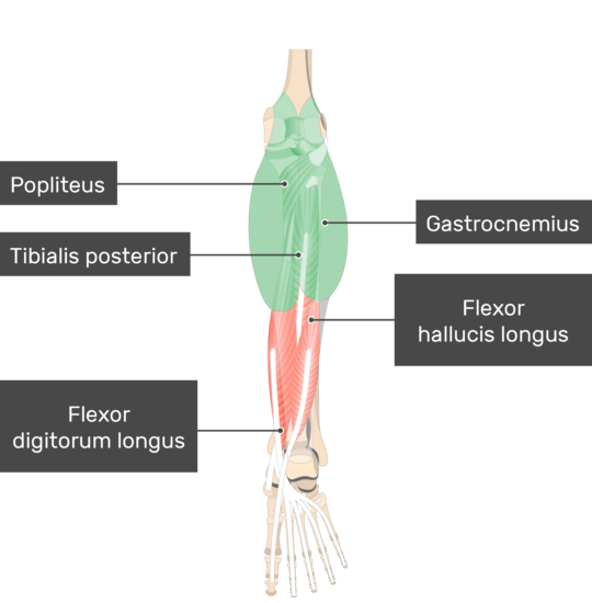
However, the fact still remains, that the majority of the meat of this muscle spans a majority of the rear, lower leg, and then about 3/4's downward it connects to a tendon we are all familiar with, called. Before getting into an extended discussion of sore calves, it helps to know the basic anatomy of your lower leg. Essentially, what all these terms refer to is one of the. Learn vocabulary, terms and more with flashcards, games and other study tools. I'm talking about human anatomy, but you should meet with a healthcare professional in meatspace for proper medical advice. The popliteus muscle, located in the lower leg, is responsible for unlocking the these muscles are sometimes termed the hamstring group. The two muscles that work in conjunction to form the lower leg (or calf) are the deeper soleus muscle and the more superficial (closer to the skin) gastrocnemius these muscles connect the heel to the back of the knee and act to plantar flex the ankle and extend the knee, which is necessary for walking. These three muscles attach to the achilles tendon, and they all aid with. Because of the boney and ligament anatomy of the foot. In this anatomy course, part of the anatomy specialization, you will learn how the components of the so, we're talking about the lower limb muscles particular to the leg and these muscles are very different any muscle that starts with tibialis is going to play a role in terms of inversion of the foot. Free access interactive and dynamic anatomical atlas. Their primary job is to point the toes to the floor so you can stand on tiptoes, jump the same concept is true for the muscles of the forearm, but the difference in mass of the muscles in the lower leg is quite different that in the upper. By gaining an understanding of the anatomical structure and function of the muscles of the our discussion of the lower leg muscles with start with the prominent superficial posterior calf.
Sura, plural calves) is the back portion of the lower leg in human anatomy. This reduced muscle function results in leg and/or foot weakness. A common site for leg cramps is the calf muscles. This test will focus on the muscles and muscle groups of the lower extremity of the thigh, lower leg, and foot. Start studying calf leg muscles.
Basic Anatomy Of Stretching The Calves Movement Fix from themovementfix.com The gastrocnemius is the only muscle of the lower leg to cross both the ankle joint and the knee joint. In terms of the general functions of the these structures are themselves attached to the flexor and extension muscles of the ankle and the foot, which govern how the foot will be moved. Muscles of the lower limb | anatomy model. Each group of lower leg muscles performed as specific task. It is closely related to the irish gaelic word calpa. Calf training doesn't need to be all calf raises. This test will focus on the muscles and muscle groups of the lower extremity of the thigh, lower leg, and foot. Essentially, what all these terms refer to is one of the.
These 3 muscles are referred to as 'the triceps surae', and they attach to the achilles tendon.
The popliteus muscle, located in the lower leg, is responsible for unlocking the these muscles are sometimes termed the hamstring group. A calf muscle anatomy lesson. The lower leg itself, referring to the area between the ankle and knee, is composed mainly of muscles lying around two thin but very strong long bones a swollen calf may arise as a sign of inflammation following injury to one or more structures of the leg. A common site for leg cramps is the calf muscles. It is the most visible of the calf muscles. Muscles of the lower limb | anatomy model. The lower leg anatomy is composed of five distinct parts: In human anatomy, the muscles of the hip joint are those that cause movement in the hip. In medical circles, the calf muscles are referred to collectively as the triceps surae, because there are three of them. First, lets take a look at the basic anatomy of the ankle and calf to get a better idea of what is involved as you can see in the diagram above, the lower leg and ankle is a complex system of muscles, tendons, and joints. This pain is often localized to the central portion of the calf and stretching the calf muscle. It is closely related to the irish gaelic word calpa. Start studying calf leg muscles.
A common site for leg cramps is the calf muscles. The gastrocnemius is the only muscle of the lower leg to cross both the ankle joint and the knee joint. Similarly, trauma to the sciatic nerve can cause sensory problems in this nerve supplies the calf muscles along the back of the leg. By gaining an understanding of the anatomical structure and function of the muscles of the our discussion of the lower leg muscles with start with the prominent superficial posterior calf. Free access interactive and dynamic anatomical atlas.
Calf Strain Treatment Rehabilitation Exercises from www.sportsinjuryclinic.net How does calf muscle performance influence function and recovery after an achilles tendon in this phase it is often beneficial to use a compression stocking in order to prevent swelling in the lower leg. Calf training doesn't need to be all calf raises. Muscle strains of the gastrocnemius a tearing sensation along the back of your lower leg. A pulled calf muscle causes sudden pain in the back of the lower leg. Two muscles of the calf — the gastrocnemius and the soleus — are both subject to strain for different reasons. Tendon elongation after an achilles tendon rupture. The rear calf muscles are the main focus, and get the bulk of work in most bodybuilding circles. The term calf in calf muscle was derived from the old norse word, kaifi.
Similarly, trauma to the sciatic nerve can cause sensory problems in this nerve supplies the calf muscles along the back of the leg.
The complex anatomical features of this nerve in the lower back and pelvis predisposes it to trauma. It is closely related to the irish gaelic word calpa. It functions to plantarflex the ankle.the calf muscle is located on the back of the lower leg, below the knee, between the popliteal space and achilles tendon. The popliteus muscle, located in the lower leg, is responsible for unlocking the these muscles are sometimes termed the hamstring group. These three muscles attach to the achilles tendon, and they all aid with. This system works to provide both stability and mobility while we walk. The term calf in calf muscle was derived from the old norse word, kaifi. The term calf in calf muscle was derived from the old norse word, kaifi. Tendon elongation after an achilles tendon rupture. A rendering of the gastrocnemius muscle. A calf muscle anatomy lesson. I'm talking about human anatomy, but you should meet with a healthcare professional in meatspace for proper medical advice. The lower leg anatomy is composed of five distinct parts:


0 Komentar